
Japanese / English
Image quality evaluation of whole slide image (WSI)
A whole slide imaging scanner (WSI) scans the pathological specimen to produce a high- quality digital image to perform analysis and diagnosis on a computer screen. However, the scanner sometimes fails to produce sufficient quality image which is the pre-condition of productive analysis and reliable diagnosis. Therefore, the image quality of WSI must be evaluated to select efficient WSI for analysis and diagnosis. In a previous study, a reference-less quality evaluation method (RQM) was proposed to evaluate regions of interest (ROI) of WSI based on sharpness and noise. [1] A reference-less evaluation method is suitable for the WSI system as the reference image is difficult to obtain. The method was found robust against both objective (MSE) and pathologists’ subjective scores. However, this method resulted in false positives for tissue-artifact regions and failed to select appropriate WSI for analysis in the presence of tissue artifacts in the slide.
The WSI quality is mainly affected by scanner failures such as focus problem or noise and slide condition such as tissue artifacts. If the quality is compromised due to scanner failure only, it is necessary to rescan the slide. When it is worthless to rescan the slide if the quality is affected by tissue artifacts as rescanning can’t recover the artifacts. Therefore, an efficient WSI system must discriminate the cause of quality failure and act accordingly to deal with the failure in the pathology workflow. Thus, we proposed a system to detect the tissue artifacts (i.e. tissue fold and air bubble) in the first step and then evaluate the image quality based on sharpness degradation and noise excluding artifacts [Fig. 1] utilizing RQM method. [2,3] After that the WSI is selected for analysis if the percentage of poor-quality regions in the WSI is lower than a threshold; otherwise the slide is recommended for rescanning. Our system detects artifacts at low magnification (1x) and then evaluates quality at high magnification (20x) to save time which is important for practical use. For artifact detection, we investigated feature selection and rotation invariance classification. This system relies on SVM classifier with optimally selected features for detecting artifact as it outperformed the CNN classifier, trained on a limited data. The proposed system enables accurate selection of slide for analysis which was
demonstrated experimentally [Fig. 2, Table 1]. Moreover, an exemplary image analysis application was developed to demonstrate the usability of the proposed system. This system is under use in the hospital and will be updated based on the feedbacks from pathologists.
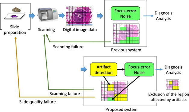
Fig 1. Proposed quality evaluation approach
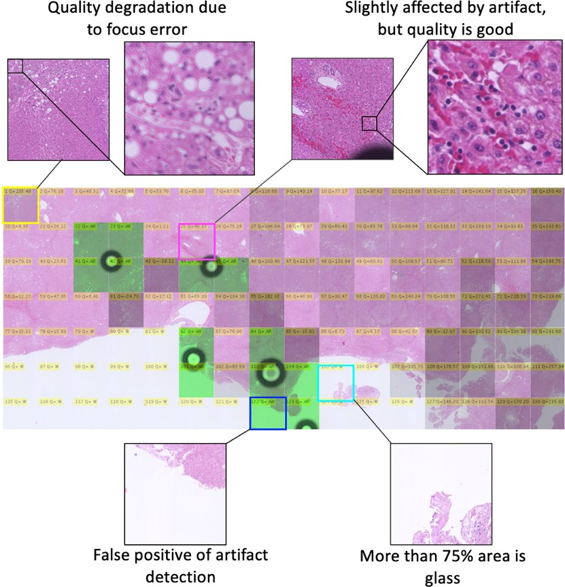
Fig. 2 Quality evaluation of typical WSI

Table 1. Comparison of proposed method with RQM
This is collaborative research between Memorial Sloan Kettering Cancer Center, USA, Shinshu University and Tokyo Institute of Technology, Japan. The author acknowledges the support (in-part) of JST-Mirai Grant Program.
Related publications
- Hashimoto Noriaki, Pinky A. Bautista, Masahiro Yamaguchi, Nagaaki Ohyama, Yukako Yagi. Referenceless image quality evaluation for whole slide imaging, Journal of Pathology Informatics, 3(1),9(2012).
- Hossain M. Shakhawat, Tomoya Nakamura, Fumikazu Kimura, Yukako Yagi, Masahiro Yamaguchi. Practical image quality evaluation for whole slide imaging scanner, The 4th Biomedical Imaging and Sensing Conference 2018 (BISC2018), Proceedings of SPIE, 10711, 107111S, Apr. 2018.
- Hossain M. Shakhawat, Tomoya Nakamura, Fumikazu Kimura, Yukako Yagi, Masahiro Yamaguchi. Automatic detection of image quality failure for the practical use of whole slide imaging scanner, JAMIT Annual Meeting 2018, 第 37 回日本医用画像工学会大会予稿集, p. 58, Jul. 2018.
- Hossain M. Shakhawat, Tomoya Nakamura, Fumikazu Kimura, Yukako Yagi, Masahiro Yamaguchi. Automatic quality evaluation of whole slide images for the practical use of whole slide imaging scanner, ITE Trans. on MTA Vol. 8, No. 4, pp. 252-268 (2020).