
Japanese / English
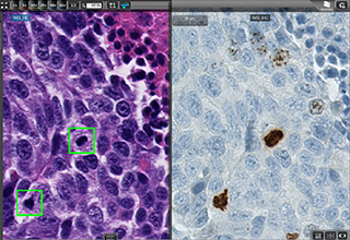
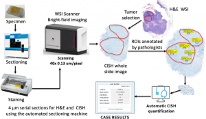
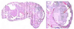
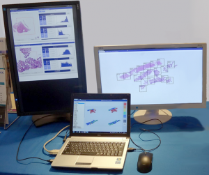
Pathology image analysis
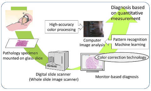
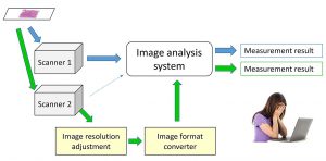
Color correction of WSI
Digital Staining using multispectral imaging
Publications
- Jiaming Li, Toshiyuki Adachi, Saori Takeyama, Masahiro Yamaguchi, and Yukako Yagi. U-Net based mitosis detection from H&E-stained images with the semi-automatic annotation using pHH3 IHC-stained images, Proc. SPIE 12032, Medical Imaging 2022: Image Processing, 120322J (4 April 2022); https://doi.org/10.1117/12.2612815
- Saori Takeyama, Tomoaki Watanabe, Masahiro Yamaguchi, Takumi Urata, Fumikazu Kimura, Keiko Ishii. Dye Amount Estimation in a Papanicolaou-stained Specimen using Multispectral Imaging, The 29th Color and Imaging Conference (CIC29), Society for Imaging Science and Technology, A-09, Nov. 2021. DOI : 10.2352/issn.2169-2629.2021.29.179
- Maulana Abdul Aziz, Tomoya Nakamura, Masahiro Yamaguchi, Tomoharu Kiyuna, Yoshiko Yamashita, Tokiya Abe, Akinori Hashiguchi, Michiie Sakamoto. Effectiveness of color correction on the quantitative analysis of histopathological images acquired by different whole-slide scanners, Artificial Life and Robotics, Jul. 2018. [SharedIt ]
- Masahiro Yamaguchi. Toward the generic quantification of morphological and textural features: the impact of image standardization, 3rd Digital Pathology Congress Asia 2017.(2017)
- Masahiro Yamaguchi. Computer-Aided Differentiation for Pathology Images, Image-Based Computer-Assisted Radiation Therapy, Springer, pp. 67-84, Mar. 2017.
- Yoshiko Yamashita, Shu Ichihara, Suzuko Moritani, Han-Seung Yoon, Masahiro Yamaguchi. Does flat epithelial atypia have rounder nuclei than columnar cell change/hyperplasia? A morphometric approach to columnar cell lesions of the breast, Virchows Archiv, Vol. 468(6), pp. 663-673. (2016)[SharedIt]
- Masahiro Ishikawa, Yuri Murakami, Sercan Taha AHI, Masahiro Yamaguchi, naoki kobayashi, Tomoharu Kiyuna, Yoshiko Yamashita, akira saito, Tokiya Abe, Akinori Hashiguchi, Michiie Sakamoto. Automatic quantification of morphological features for hepatic trabeculae analysis in stained liver specimens., Journal of Medical Imaging, Vol. 3, No. 2, 027502. (2016)
- Maulana Abdul Aziz, Hiroshi Kanazawa, Yuri Murakami, Fumikazu Kimura, Masahiro Yamaguchi, Tomoharu Kiyuna, Yoshiko Yamashita, Akira Saito, Masahiro Ishikawa, Naoki Kobayashi, Tokiya Abe, Akinori Hashiguchi, Michiie Sakamoto. Enhancing automatic classification of hepatocellular carcinoma images through image masking, tissue changes and trabecular features, Journal of Pathology Informatics, Vol. 6, No. 1, 26.(2015)
- Masahiro Ishikawa, Naoki Kobayashi, Hideki Komagata, Kazuma Shinoda, Masahiro Yamaguchi, Tokiya Abe, Akinori Hashiguchi, Michiie Sakamoto. An accurate method of extracting fat droplets in liver images for quantitative evaluation, SPIE Medical Imaging 2015: Digital Pathology, Medical Imaging 2015: Digital Pathology, Proc. SPIE, SPIE: International Society for Optics and Photonics, Vol. 9420. (2015)
- Yuri Murakami, Tokiya Abe, Akinori Hashiguchi, Masahiro Yamaguchi, Michiie Sakamoto. Color correction for automatic fibrosis quantification in liver biopsy specimens, Journal of Pathology Informatics, 4:36. (2013)
- N. Hashimoto, P. A. Bautista, M. Yamaguchi, N. Ohyama, Y. Yagi, “Referenceless image quality evaluation for whole slide imaging,” Journal of Pathology Informatics, Vol. 3, Mar. (2012)[PDF]
- M. Tashiro, Y. Murakami, T. Obi, M. Yamaguchi, N. Ohyama, “Layered scalable coding of multispectral images based on visible component separation,” Optical Review, Vol. 18, No. 6, pp. 462-469, Nov. (2011)
- N. Hashimoto, Y. Murakami, P. A. Bautista, M. Yamaguchi, T. Obi, N. Ohyama, K. Uto, Y. Kosugi, “Multispectral image enhancement for effective visualization,” Optics Express, Optical Society of America, vol. 19, no. 10, pp. 9315-9329, Apr. (2011)
- C. Atupelage, H. Nagahashi, M. Yamaguchi, M. Sakamoto, A. Hashiguchi, “Multifractal Feature Based Cancer Detection for Pathological Images,” The 5th International Conference on Bioinformatics and Biomedical Engineering, CDROM Proceeding of ICBBE’2011, May. (2011)
- M. Tashiro, R. Yoshida, T. Miyazawa, Y. Murakami, M. Yamaguchi, N. Ohyama, Y. Yagi. “H&E Digital Staining From the Multispectral Image of a Specimen Stained With Hematoxylin Only,” APIII 2009 Advancing Practice, Instruction, & Innovation through Informatics. Abstract ID – 744. (2009)
- P. A. Bautista, T. Abe, M. Yamaguchi, Y. Yagi and N. Ohyama, “Multispectral image enhancement for H&E stained pathological tissue specimens,” Proc. SPIE, Vol. 6918, 691836 (2008)
- P. A. Bautista, T. Abe, M. Yamaguchi, Y. Yagi and N. Ohyama, “Digital staining of pathological tissue specimens using spectral transmittance,” Proc. SPIE, 5747, 1892-1903 (2005)
- T. Abe, Y. Murakami, M. Yamaguchi, N. Ohyama, and Y. Yagi, “Color correction of pathological images based on dye amount quantification,” Opt. Rev., 12, 4, 293-300, (2005)[SharedIt]
- P. Bautista, T. Abe, M. Yamaguchi, Y. Yagi, and N. Ohyama, “Digital staining for multispectral images of pathological tissue specimens based on combined classification of spectral transmittance,” Comput. Med. Imaging Graph. 29, 649-657, (2005)[SharedIt]
- T. Abe, M. Yamaguchi, Y. Murakami, N. Ohyama,Y. Yagi, “Color correction of pathological images for different staining-condition slides,” IEEE HealthCom 2004, Odawara, Japan, 218-223, (2004)
- H.Fukuda, N.Ohyama, M.Yamaguchi and T.Wada, Development of Multi-Spectral Imaging System for Pathology, Advancing Pathology Informatics, Imaging, and the Internet (APIII 2002),Pittsburgh, PA, 2002
- K. Fujii, M. Yamaguchi, N. Ohyama, K. Mukai, “Development of support system for pathology using spectral transmittance -the quantification method of stain conditions,” Proc SPIE, Medical Imaging, vol.4684, 1516-1523 (2002)