
Japanese / English
Automated analysis of whole slide image (WSI)
The automated WSI analysis system is applied to histopathology diagnosis of hepatocellular carcinoma [Fig. 1]. The overview of the system is shown in fig. 2. After preprocessing such as color correction, the nuclei detection and nuclear feature measurement are employed. Also, stromal areas, sinusoids and fat droplets are segmented for the structural feature measurements. The system shows the quantification results as a heat map. Moreover, the cancer discrimination is performed by SVM (support vector machine) using the feature vector composed of nuclei and structural features. The result of feature measurement and SVM classification are also visualized as a heat map as shown in fig. 3. The accuracy of classification was tested by 5-fold cross-validation using about 1,000 ROIs. As a result, both the sensitivity and specificity were almost 90% [Table 1]. The heat map of the possibility of malignancy will support pathologists to pick up the regions that should be observed carefully, and the heat map of each feature provides pathologists supportive information for subtype classification, grading, treatment selection, and reasoning of the diagnosis.
Demonstration at IADP 2014 / CIC22 (2014, Boston)
[Link https://www.oid.ict.e.titech.ac.jp/wp/home_en/research/pathology/automated-analysis/iadp-2014-cic22/ ]
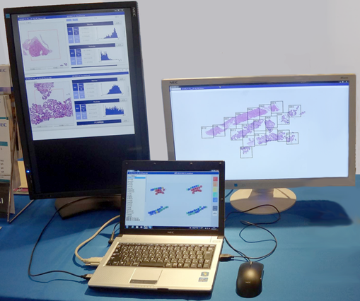
Fig. 1 Prototype system

Fig. 2 Flow diagram of the prototype system

Fig. 3 Heat map for HCC probability
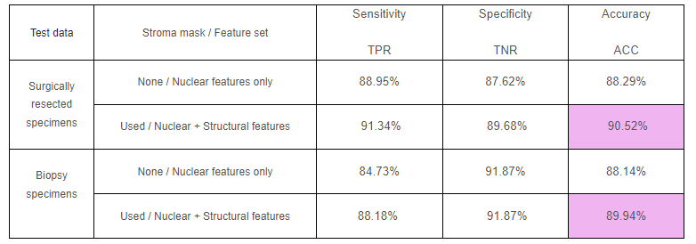
Table 1 Classification result
The research project, “image analysis technology for quantitative pathological diagnosis” was carried out by Keio University, NEC Corporation, Saitama Medical University and Tokyo Institute of Technology as a commissioned research by NEDO (New Energy and Industrial Technology Development Organization). I acknowledge Professor Michiie Sakamoto, Dr. Akinori Hashiguchi, and Dr. Tokiya Abe, Keio University, Dr. Akira Saito, Dr. Kenichi Kamijo, Dr. Tomoharu Kiyuna, Kamei, Dr. Yoshiko Yamashita, NEC, Professor Naoki Kobayashi and Dr. Masahiro Ishikawa, Saitama Medical School, Dr. Yuri Murakami and Professor Hiroshi Nagahashi, formerly Tokyo Institute of Technology, Dr. Hiroshi Kanazawa, Dr. Fumio Kimura, and the students in Optical Imaging and Display group, Tokyo Institute of Technology.
References
- M. Yamaguchi. Computer-Aided Differentiation for Pathology Images, Image-Based Computer-Assisted Radiation Therapy, Springer, pp. 67-84, Mar. 2017.
- M. Ishikawa, Y. Murakami, S. T. AHI, M. Yamaguchi, N. Kobayashi, T. Kiyuna, Y. Yamashita, A. Saito, T. Abe, A. Hashiguchi, M. Sakamoto, “Automatic quantification of morphological features for hepatic trabeculae analysis in stained liver specimens,” Journal of Medical Imaging, Vol. 3, No. 2, 027502, Jun. 2016.
- M. A. Aziz, H. Kanazawa, Y. Murakami, F. Kimura, M. Yamaguchi, T. Kiyuna, Y. Yamashita, A. Saito, M. Ishikawa, N. Kobayashi, T. Abe, A. Hashiguchi, M. Sakamoto, “Enhancing automatic classification of hepatocellular carcinoma images through image masking, tissue changes and trabecular features,” Journal of Pathology Informatics, Vol. 6, No. 1, 26, Jun. 2015.
- K. Shinoda, N. Kobayashi, A. Katoh, H. Komagata, M. Ishikawa, Y. Murakami, M. Yamaguchi, T. Abe, A. Hashiguchi, M. Sakamoto, “An Efficient Wavelet-Based ROI Coding for Multiple Regions,” IEICE Transactions on Fundamentals of Electronics, Communications and Computer Sciences, Vol. E98.A, No. 4, pp. 1006-1020, Apr. 2015.
- M. Ishikawa, N. Kobayashi, H. Komagata, K. Shinoda, M. Yamaguchi, T. Abe, A. Hashiguchi, M. Sakamoto, “An accurate method of extracting fat droplets in liver images for quantitative evaluation,” SPIE Medical Imaging 2015: Digital Pathology, Proc. SPIE, Vol. 9420, Mar. 2015.
- C. Atupelage, H. Nagahashi, F. Kimura, M. Yamaguchi, A. Tokiya, A. Hashiguchi, M. Sakamoto, “Computational hepatocellular carcinoma tumor grading based on cell nuclei classification,” Journal of Medical Imaging, Vol. 1, No. 3, pp. 034501, Oct. 2014.
- T. Abe, Y. Murakami, M. Yamaguchi, Y. Yamashita, T. Kiyuna, K. Yamazaki, A. Hashiguchi, Y. Yasui, M. Kurosaki, N. Izumi, M. Sakamoto, “Whole Slide Image Analysis System for Quantification of Liver Fibrosis,” 2nd International Congress of the International Academy of Digital Pathology (IADP), Analytical Cellular Pathology, Vol. 2014, Article ID 505968, Nov. 2014.
- Y. Murakami, T. Abe, Y. Yamashita, M. Yamaguchi, M. Ishikawa, A. Hashiguchi, T. Kiyuna, A. Saito, M. Sakamoto, “Color processing in pathology image analysis system for liver biopsy,” Proc. Color and Imaging Conference 2014, pp. 184-188, Nov. 2014.
- Y. Yamashita, T. Kiyuna, M. Sakamoto, A. Hashiguchi, M. Ishikawa, Y. Murakami, M. Yamaguchi, “Development of a prototype for hepatocellular carcinoma classification based on morphological features automatically measured in whole slide images,” 2nd International Congress of the International Academy of Digital Pathology (IADP), Analytical Cellular Pathology, Vol. 2014, Article ID 817192, Nov. 2014.
- C. Atupelage, H. Nagahashi, M. Yamaguchi, F. Kimura, T. Abe, A. Hashiguchi, M. Sakamoto, “Cell Nuclei Classification in HE-stained Biopsy Images,” Proc. 2014 Int. Conf. Biology and Biomedical Engineering (BBE’14), pp. 55-60, Mar. 2014.
- Y. Murakami, T. Abe, A. Hashiguchi, M. Yamaguchi, M. Sakamoto, “Color correction for automatic fibrosis quantification in liver biopsy specimens,” Journal of Pathology Informatics, 4:36. (2013)
- C. Atupelage, H. Nagahashi, M. Yamaguchi, T. Abe, A. Hashiguchi, M. Sakamoto, “Computational grading of hepatocellular carcinoma using multifractal feature description,” Computerized Medical Imaging and Graphics, Vol. 37, No. 1, pp. 61-71, Jan. 2013.
- M. Ishikawa, S. T. Ahi, Y. Murakami, F. Kimura, M. Yamaguchi, T. Abe, A. Hashiguchi, M. Sakamoto, “Automatic segmentation of hepatocellular structure from HE-stained liver tissue,” SPIE Medical Imaging 2013: Digital Pathology, Proc. SPIE, Vol. 8676, 867611, Mar. 2013.
- C. Atupelage, H. Nagahashi, M. Yamaguchi, T. Abe, A. Hashiguchi, M. Sakamoto, “Classification of prostate histopathology images based on multifractal analysis,” IEICE Transaction on Information Processing, Vol. E95-D, No. 12, pp. 3037-3045, Dec. 2012.
- C. Atupelage, H. Nagahashi, M. Yamaguchi, T. Abe, A. Hashiguchi, M. Sakamoto, “Multifractal Feature Descriptor for Grading Hepatocellular Carcinoma,” Proc. 21th International Conference on Pattern Recognition (ICPR’2012), pp. 129-132, Nov. 2012.
- C. Atupelage, H. Nagahashi, M. Yamaguchi, M. Sakamoto, A. Hashiguchi, “Multifractal Feature Descriptor for Histopathology,” The 1st Congress of the International Academy of Digital Pathology, Analytical Cellular Pathology, Volume 35, Issue 2, Pages 123-126, Aug. 2011.