
Japanese / English
Image analysis technology for quantitative pathology
Topics of research
- Automated analysis of whole slide image (WSI)
- Image segmentation for extraction tissue elements
- Accuracy enhancement of color and spectral information
- Color correction of WSI
- Application of multispectral imaging technique
- Digital Staining
Fig.1 16-band Multispectral Microscopic Imaging System
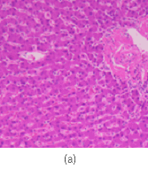
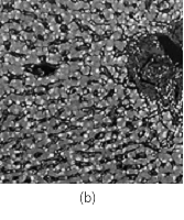
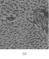
Fig.2 (a) HE stained image, (b) Unmixed Hematoxylin image, (c) Unmixed Eosin image
Fig.3 Standardization of staining condition using digital image processing
Fig.4 HE image Digital staining for fibrosis visualization Real MT stain
Publications
- Masahiro Yamaguchi. Toward the generic quantification of morphological and textural features: the impact of image standardization, 3rd Digital Pathology Congress Asia 2017.(2017)
- Yoshiko Yamashita, Shu Ichihara, Suzuko Moritani, Han-Seung Yoon, Masahiro Yamaguchi. Does flat epithelial atypia have rounder nuclei than columnar cell change/hyperplasia? A morphometric approach to columnar cell lesions of the breast, Virchows Archiv, Vol. 468(6), pp. 663-673. (2016)
- Masahiro Ishikawa, Yuri Murakami, Sercan Taha AHI, Masahiro Yamaguchi, naoki kobayashi, Tomoharu Kiyuna, Yoshiko Yamashita, akira saito, Tokiya Abe, Akinori Hashiguchi, Michiie Sakamoto. Automatic quantification of morphological features for hepatic trabeculae analysis in stained liver specimens., Journal of Medical Imaging, Vol. 3, No. 2, 027502. (2016)
- Maulana Abdul Aziz, Hiroshi Kanazawa, Yuri Murakami, Fumikazu Kimura, Masahiro Yamaguchi, Tomoharu Kiyuna, Yoshiko Yamashita, Akira Saito, Masahiro Ishikawa, Naoki Kobayashi, Tokiya Abe, Akinori Hashiguchi, Michiie Sakamoto. Enhancing automatic classification of hepatocellular carcinoma images through image masking, tissue changes and trabecular features, Journal of Pathology Informatics, Vol. 6, No. 1, 26.(2015)
- 山下慶子, 喜友名朝春, 山口雅浩. 乳腺Ki-67計測定量化のための全自動画像解析システムの開発, Medical Imaging Technology, Vol. 33, No. 3, pp. 124-132. (2015)
- Masahiro Ishikawa, Naoki Kobayashi, Hideki Komagata, Kazuma Shinoda, Masahiro Yamaguchi, Tokiya Abe, Akinori Hashiguchi, Michiie Sakamoto. An accurate method of extracting fat droplets in liver images for quantitative evaluation, SPIE Medical Imaging 2015: Digital Pathology, Medical Imaging 2015: Digital Pathology, Proc. SPIE, SPIE: International Society for Optics and Photonics, Vol. 9420. (2015)
- Masahiro Ishikawa, Sercan Taha Ahi, Fumikazu Kimura, Masahiro Yamaguchi, Hiroshi Nagahashi, Akinori Hashiguchi, Michiie Sakamoto. Segmentation of Sinusoids in Hematoxylin and Eosin Stained Liver Specimens Using an Orientation-Selective Filter, Open Journal of Medical Imaging, Vol. 3, No. 4, pp. 144-155. (2013)
- Yuri Murakami, Tokiya Abe, Akinori Hashiguchi, Masahiro Yamaguchi, Michiie Sakamoto. Color correction for automatic fibrosis quantification in liver biopsy specimens, Journal of Pathology Informatics, 4:36. (2013)
- N. Hashimoto, P. A. Bautista, M. Yamaguchi, N. Ohyama, Y. Yagi, “Referenceless image quality evaluation for whole slide imaging,” Journal of Pathology Informatics, Vol. 3, Mar. (2012)
- M. Tashiro, Y. Murakami, T. Obi, M. Yamaguchi, N. Ohyama, “Layered scalable coding of multispectral images based on visible component separation,” Optical Review, Vol. 18, No. 6, pp. 462-469, Nov. (2011)
- N. Hashimoto, Y. Murakami, P. A. Bautista, M. Yamaguchi, T. Obi, N. Ohyama, K. Uto, Y. Kosugi, “Multispectral image enhancement for effective visualization,” Optics Express, Optical Society of America, vol. 19, no. 10, pp. 9315-9329, Apr. (2011)
- C. Atupelage, H. Nagahashi, M. Yamaguchi, M. Sakamoto, A. Hashiguchi, “Multifractal Feature Based Cancer Detection for Pathological Images,” The 5th International Conference on Bioinformatics and Biomedical Engineering, CDROM Proceeding of ICBBE’2011, May. (2011)
- M. Tashiro, R. Yoshida, T. Miyazawa, Y. Murakami, M. Yamaguchi, N. Ohyama, Y. Yagi. “H&E Digital Staining From the Multispectral Image of a Specimen Stained With Hematoxylin Only,” APIII 2009 Advancing Practice, Instruction, & Innovation through Informatics. Abstract ID – 744. (2009)
- P. A. Bautista, T. Abe, M. Yamaguchi, Y. Yagi and N. Ohyama, “Multispectral image enhancement for H&E stained pathological tissue specimens,” Proc. SPIE, Vol. 6918, 691836 (2008)
- P. A. Bautista, T. Abe, M. Yamaguchi, Y. Yagi and N. Ohyama, “Digital staining of pathological tissue specimens using spectral transmittance,” Proc. SPIE, 5747, 1892-1903 (2005)
- T. Abe, Y. Murakami, M. Yamaguchi, N. Ohyama, and Y. Yagi, “Color correction of pathological images based on dye amount quantification,” Opt. Rev., 12, 4, 293-300, (2005)
- P. Bautista, T. Abe, M. Yamaguchi, Y. Yagi, and N. Ohyama, “Digital staining for multispectral images of pathological tissue specimens based on combined classification of spectral transmittance,” Comput. Med. Imaging Graph. 29, 649-657, (2005)
- P. Bautista, T. Abe, M. Yamaguchi, et al.,” Digital staining of unstained pathological tissue samples through spectral transmittance classification”, Optical Review, vol.12, 7-14, (2005)
- T. Abe, M. Yamaguchi, Y. Murakami, N. Ohyama,Y. Yagi, “Color correction of pathological images for different staining-condition slides,” IEEE HealthCom 2004, Odawara, Japan, 218-223, (2004)
- H.Fukuda, N.Ohyama, M.Yamaguchi and T.Wada, “Development of Multi-Spectral Imaging System for Pathology”, Advancing Pathology Informatics, Imaging, and the Internet (APIII 2002), PA, 2002
- K. Fujii, M. Yamaguchi,, N. Ohyama, K. Mukai, “Development of support system for pathology using spectral transmittance -the quantification method of stain conditions,” Proc SPIE, Medical Imaging, vol.4684, 1516-1523 (2002)