
Japanese / English
「定量的病理診断」のための画像解析技術
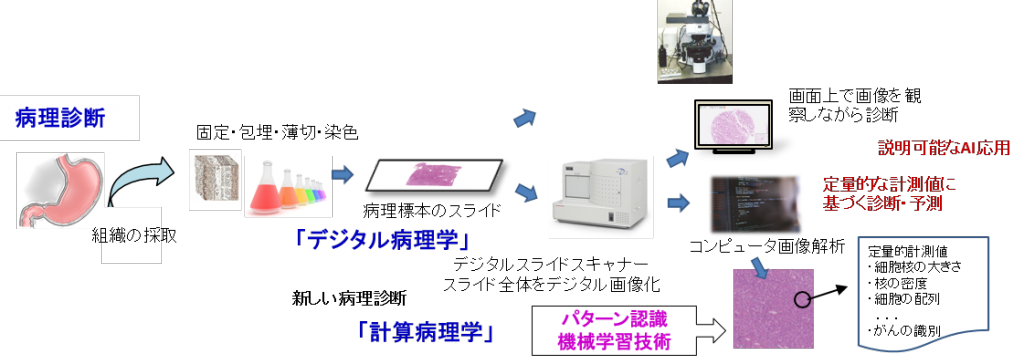
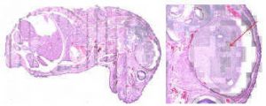
病理診断は、病変部の組織を顕微鏡で観察して良性/悪性の判定や進行度等を判定するもので、がん等の治療方針を決めるにあたって重要な役割を果たします。本研究では、「デジタルスライド」技術に基づいて得られるデジタル病理画像に対してパターン解析技術を適用し、「定量的」「高精度」な病理診断の実現を目指しています。
研究テーマ
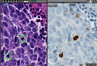
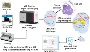
•深層学習を用いた細胞や組織構造などの画像パターン認識技術
•画像解析による診断に有用な指標の算出
•病理画像解析のための色補正技術
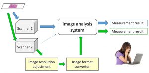
普遍的な特徴量取得のための画像標準化
病理全スライド画像(Whole Slide Image)の画質評価
病理画像の色標準化技術
Publications
- Jiaming Li, Toshiyuki Adachi, Saori Takeyama, Masahiro Yamaguchi, and Yukako Yagi. U-Net based mitosis detection from H&E-stained images with the semi-automatic annotation using pHH3 IHC-stained images, Proc. SPIE 12032, Medical Imaging 2022: Image Processing, 120322J (4 April 2022); https://doi.org/10.1117/12.2612815
- Masahiro Yamaguchi. Toward the generic quantification of morphological and textural features: the impact of image standardization, 3rd Digital Pathology Congress Asia 2017.(2017)
- Yoshiko Yamashita, Shu Ichihara, Suzuko Moritani, Han-Seung Yoon, Masahiro Yamaguchi. Does flat epithelial atypia have rounder nuclei than columnar cell change/hyperplasia? A morphometric approach to columnar cell lesions of the breast, Virchows Archiv, Vol. 468(6), pp. 663-673. (2016)[SharedIt]
- Masahiro Ishikawa, Yuri Murakami, Sercan Taha AHI, Masahiro Yamaguchi, naoki kobayashi, Tomoharu Kiyuna, Yoshiko Yamashita, akira saito, Tokiya Abe, Akinori Hashiguchi, Michiie Sakamoto. Automatic quantification of morphological features for hepatic trabeculae analysis in stained liver specimens., Journal of Medical Imaging, Vol. 3, No. 2, 027502. (2016)
- Maulana Abdul Aziz, Hiroshi Kanazawa, Yuri Murakami, Fumikazu Kimura, Masahiro Yamaguchi, Tomoharu Kiyuna, Yoshiko Yamashita, Akira Saito, Masahiro Ishikawa, Naoki Kobayashi, Tokiya Abe, Akinori Hashiguchi, Michiie Sakamoto. Enhancing automatic classification of hepatocellular carcinoma images through image masking, tissue changes and trabecular features, Journal of Pathology Informatics, Vol. 6, No. 1, 26.(2015)
- 山下慶子, 喜友名朝春, 山口雅浩. 乳腺Ki-67計測定量化のための全自動画像解析システムの開発, Medical Imaging Technology, Vol. 33, No. 3, pp. 124-132. (2015)
- Masahiro Ishikawa, Naoki Kobayashi, Hideki Komagata, Kazuma Shinoda, Masahiro Yamaguchi, Tokiya Abe, Akinori Hashiguchi, Michiie Sakamoto. An accurate method of extracting fat droplets in liver images for quantitative evaluation, SPIE Medical Imaging 2015: Digital Pathology, Medical Imaging 2015: Digital Pathology, Proc. SPIE, SPIE: International Society for Optics and Photonics, Vol. 9420. (2015)
- Masahiro Ishikawa, Sercan Taha Ahi, Fumikazu Kimura, Masahiro Yamaguchi, Hiroshi Nagahashi, Akinori Hashiguchi, Michiie Sakamoto. Segmentation of Sinusoids in Hematoxylin and Eosin Stained Liver Specimens Using an Orientation-Selective Filter, Open Journal of Medical Imaging, Vol. 3, No. 4, pp. 144-155. (2013)
- Yuri Murakami, Tokiya Abe, Akinori Hashiguchi, Masahiro Yamaguchi, Michiie Sakamoto. Color correction for automatic fibrosis quantification in liver biopsy specimens, Journal of Pathology Informatics, 4:36. (2013)
- N. Hashimoto, P. A. Bautista, M. Yamaguchi, N. Ohyama, Y. Yagi, “Referenceless image quality evaluation for whole slide imaging,” Journal of Pathology Informatics, Vol. 3, Mar. (2012)
- M. Tashiro, Y. Murakami, T. Obi, M. Yamaguchi, N. Ohyama, “Layered scalable coding of multispectral images based on visible component separation,” Optical Review, Vol. 18, No. 6, pp. 462-469, Nov. (2011)
- N. Hashimoto, Y. Murakami, P. A. Bautista, M. Yamaguchi, T. Obi, N. Ohyama, K. Uto, Y. Kosugi, “Multispectral image enhancement for effective visualization,” Optics Express, Optical Society of America, vol. 19, no. 10, pp. 9315-9329, Apr. (2011)
- C. Atupelage, H. Nagahashi, M. Yamaguchi, M. Sakamoto, A. Hashiguchi, “Multifractal Feature Based Cancer Detection for Pathological Images,” The 5th International Conference on Bioinformatics and Biomedical Engineering, CDROM Proceeding of ICBBE’2011, May. (2011)
- M. Tashiro, R. Yoshida, T. Miyazawa, Y. Murakami, M. Yamaguchi, N. Ohyama, Y. Yagi. H&E Digital Staining From the Multispectral Image of a Specimen Stained With Hematoxylin Only. APIII 2009 Advancing Practice, Instruction, & Innovation through Informatics. Abstract ID – 744. (2009)
- P. A. Bautista, T. Abe, M. Yamaguchi, Y. Yagi and N. Ohyama, “Multispectral image enhancement for H&E stained pathological tissue specimens,” Proc. SPIE, Vol. 6918, 691836 (2008)
- 阿部時也, 村上百合, 山口雅浩, 大山永昭, 八木由香子, “マルチスペクトル画像を用いた病理画像の色素量の定量化と色標準化-バンド数と計算精度の検討-,” MEDICAL IMAGING TECHNOLOGY. Vol. 24. No. 1. 38-47. (2006)
- P. A. Bautista, T. Abe, M. Yamaguchi, Y. Yagi and N. Ohyama, “Digital staining of pathological tissue specimens using spectral transmittance,” Proc. SPIE, 5747, 1892-1903 (2005)
- T. Abe, Y. Murakami, M. Yamaguchi, N. Ohyama, and Y. Yagi, “Color correction of pathological images based on dye amount quantification,” Opt. Rev., 12, 4, 293-300, (2005)[SharedIt]
- P. Bautista, T. Abe, M. Yamaguchi, Y. Yagi, and N. Ohyama, “Digital staining for multispectral images of pathological tissue specimens based on combined classification of spectral transmittance,” Comput. Med. Imaging Graph. 29, 649-657, (2005)
- P. Bautista, T. Abe, M. Yamaguchi, et al.,” Digital staining of unstained pathological tissue samples through spectral transmittance classification”, Optical Review, vol.12, 1-8, (2005)[SharedIt]
- T. Abe, M. Yamaguchi, Y. Murakami, N. Ohyama,Y. Yagi, “Color correction of pathological images for different staining-condition slides,” IEEE HealthCom 2004, Odawara, Japan, 218-223, (2004)
- H.Fukuda, N.Ohyama, M.Yamaguchi and T.Wada, “Development of Multi-Spectral Imaging System for Pathology”, Advancing Pathology Informatics, Imaging, and the Internet (APIII 2002), Pittsburgh, PA, 2002
- K. Fujii, M. Yamaguchi,, N. Ohyama, K. Mukai, “Development of support system for pathology using spectral transmittance -the quantification method of stain conditions,” Proc SPIE, Medical Imaging, vol.4684, 1516-1523 (2002)